


Traceable, high-quality primary cell portfolio bridges the gap between in-vitro models and in-vivo knee-joint pathophysiology
HUSTON, TX, UNITED STATES, September 2, 2026 /EINPresswire.com/ -- Knee disorders represent a major global public-health burden. Osteoarthritis, rheumatoid arthritis, meniscus injury and synovitis severely impair patient mobility and quality of life. A persistent bottleneck hindering translational progress lies in the discrepancy between conventional in-vitro cellular models and genuine in-vivo tissue conditions. Cloud-Clone delivers a comprehensive portfolio of source-quality-controlled primary cells covering four key knee-joint cell populations. These physiologically-relevant cellular resources help researchers build faithful experimental models to advance mechanistic exploration, drug screening and regenerative-medicine development for knee-joint diseases worldwide.
The knee joint is one of the human body’s most complex and weight-bearing joints, and it is highly susceptible to osteoarthritis, rheumatoid arthritis, meniscus injuries, synovitis and other pathological conditions. Statistics show that more than 500 million people worldwide live with osteoarthritis, with the knee being the most frequently affected site; osteoarthritis ranks highest among musculoskeletal disorders in terms of disability rates. Nevertheless, knee-joint disease research has long faced a fundamental challenge: the functional mismatch between in-vitro experimental models and authentic in-vivo biological status. Many interventions that show promising cellular-level effects frequently fail when translated into clinical applications.
The solution starts from the fundamental building-blocks of knee-joint tissues. Recent advances in single-cell RNA sequencing reveal that knee structures including cartilage, synovium, meniscus and ligaments encompass 10-30 distinct cellular subpopulations, each playing divergent functional roles during disease progression. Therefore, constructing reliable in-vitro research models hinges on high-quality primary cells that faithfully recapitulate native in-vivo cellular biological features.
Articular Chondrocytes: Primary Players in Cartilage Degeneration Research
Articular cartilage forms the core structure enabling smooth joint movement and load absorption, and its destruction constitutes the hallmark pathological feature of osteoarthritis. Chondrocytes are the sole resident cell type within articular cartilage, responsible for synthesising and maintaining the extracellular matrix composed of type-II collagen and proteoglycans. Latest single-cell studies have identified multiple novel chondrocyte subpopulations within osteoarthritic cartilage, including effector chondrocytes, regulatory chondrocytes, reparative chondrocytes, fibrocartilage-like chondrocytes and ferroptotic chondrocyte clusters, illustrating rich cellular heterogeneity throughout cartilage degeneration.
Primary articular chondrocytes retain donor-derived phenotypic characteristics and can be utilised to establish in-vitro degeneration models induced by IL-1β, D-galactose-triggered senescence and other stimuli. They serve as the gold-standard model for studying chondrocyte apoptosis, senescence and ferroptosis, as well as screening chondroprotective agents. Using primary chondrocyte platforms, researchers can directly observe how candidate compounds modulate type-II collagen synthesis and secretion of matrix metalloproteinases (MMP-1, MMP-13), generating robust in-vitro evidence for targeted osteoarthritis therapies.
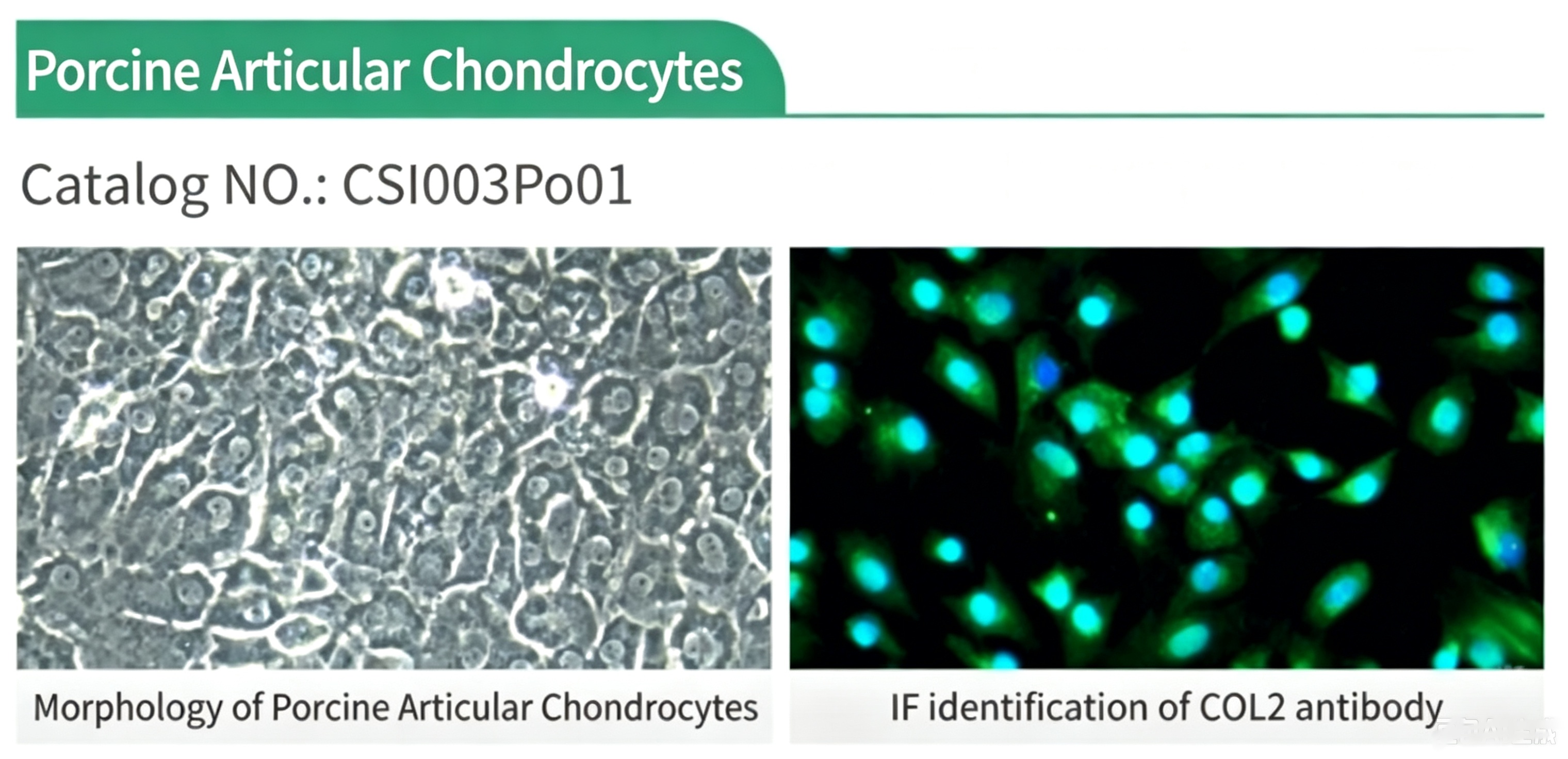
Figure 1. Morphology and COL2-based immunofluorescence identification of porcine articular chondrocytes (Cat.No. CSI003Po01)
Synovial Cells and Synovial-Derived Mesenchymal Stem Cells: Dual Roles in Inflammation Regulation and Tissue Repair
The synovium is a thin membrane (50-100 μm in thickness) lining intra-articular surfaces and is critical for joint homeostasis. Synovial-like fibroblasts (FLSs) represent the predominant cell population within synovial tissue; their aberrant activation drives synovial inflammation in rheumatoid arthritis and late-stage osteoarthritis. Research demonstrates that inflammatory signalling cascades within synovial cells (e.g. αVβ3/FAK/ERK/C-Jun pathways) can be modulated by mechanical stress, further regulating MMP secretion and cytoskeleton remodelling. Primary synovial cells constitute direct research tools for dissecting synovitis molecular mechanisms and screening anti-inflammatory therapeutics.
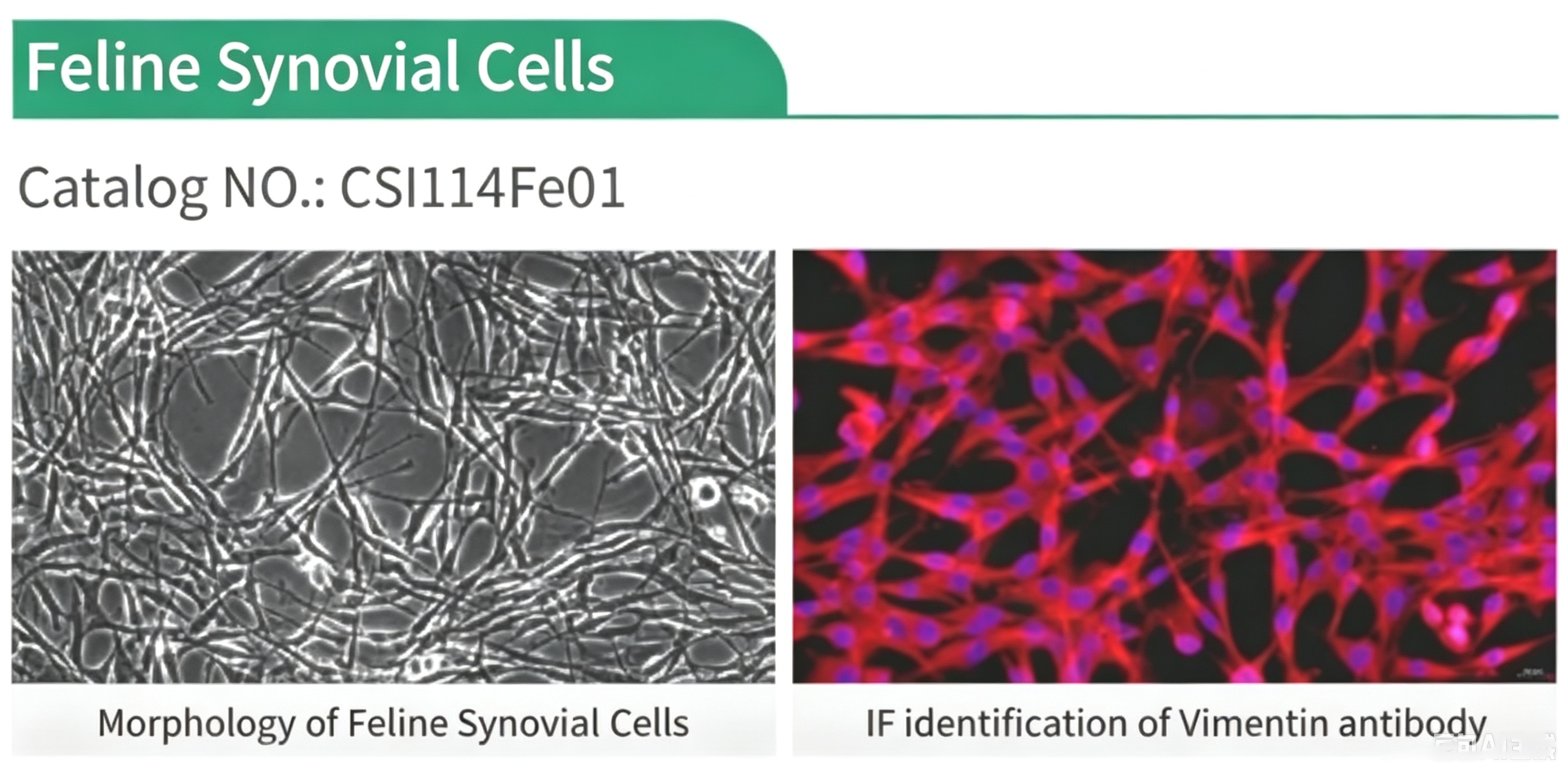
Figure 2. Morphology and vimentin-based immunofluorescence identification of feline synovial cells (Cat.No. CSI114Fe01)
Meanwhile, synovial tissue acts as an important source of mesenchymal stem cells. Studies confirm that transplanted synovial mesenchymal stem cells can differentiate into meniscal fibrocartilage cells at meniscal defect sites and interact with chondrocytes via extracellular vesicles to synergistically facilitate tissue repair. These properties render synovial-derived mesenchymal stem cells promising seed cells for regenerative medicine and tissue-engineering research, with distinctive application prospects for meniscus-injury repair and cartilage-defect regeneration.
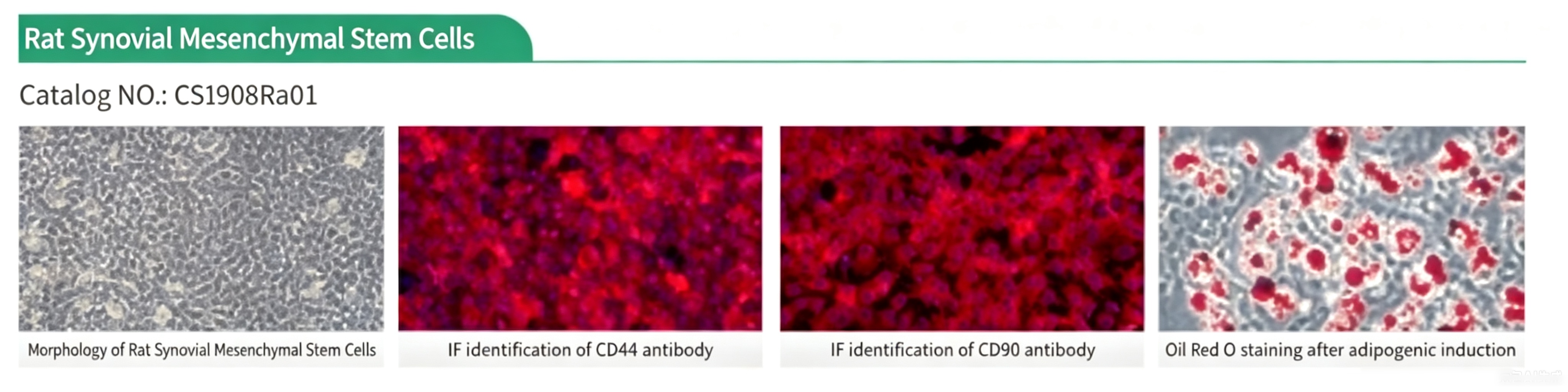
Figure 3. Morphology, immunofluorescence and histological staining of rat primary Synovial Mesenchymal Stem Cells (SMSC) (Cat.No. CSI908Ra01)
Meniscus Fibrochondrocytes: Key Guardians for Load-Bearing and Shock Absorption
Composed of fibrocartilage, the meniscus undertakes load transmission, joint stabilisation and shock-absorbing functions. The avascular inner “white zone” of the meniscus exhibits minimal self-healing capacity after injury. Meniscus fibrochondrocytes synthesise mixed matrices of type-I and type-II collagen to sustain the unique mechanical properties of meniscal tissue. Studies indicate that successful meniscus defect repair heavily relies on the capacity of transplanted cells to differentiate into meniscus-like fibrochondrocytes and survive long-term (minimum 16 weeks). Primary meniscus fibrochondrocytes serve as irreplaceable experimental models for investigating meniscus-injury repair mechanisms and screening pro-repair agents.
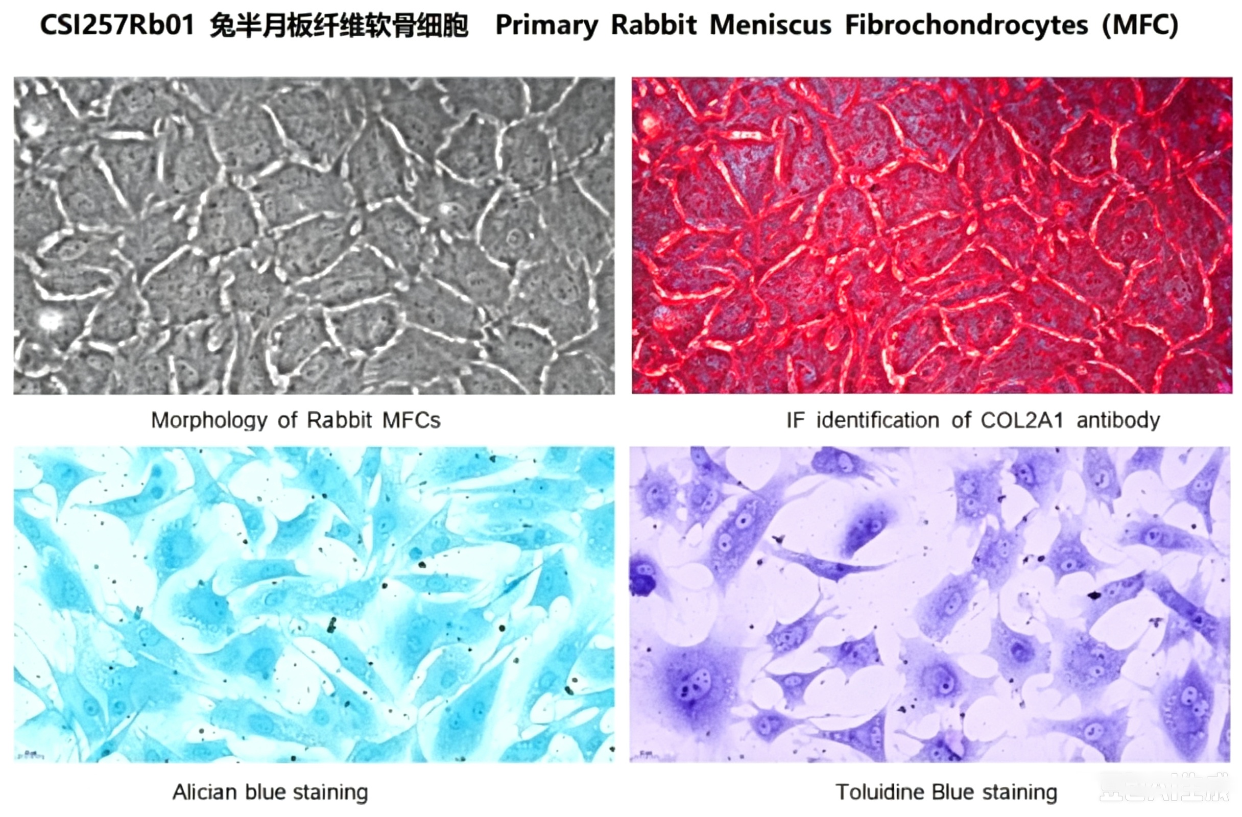
Figure 4. Morphology, immunofluorescence and histological staining of rabbit primary meniscus fibrochondrocytes (Cat.No. CSI257Rb01)
Tendon Cells: Mechanical Core for Knee-Joint Stabilising Structures
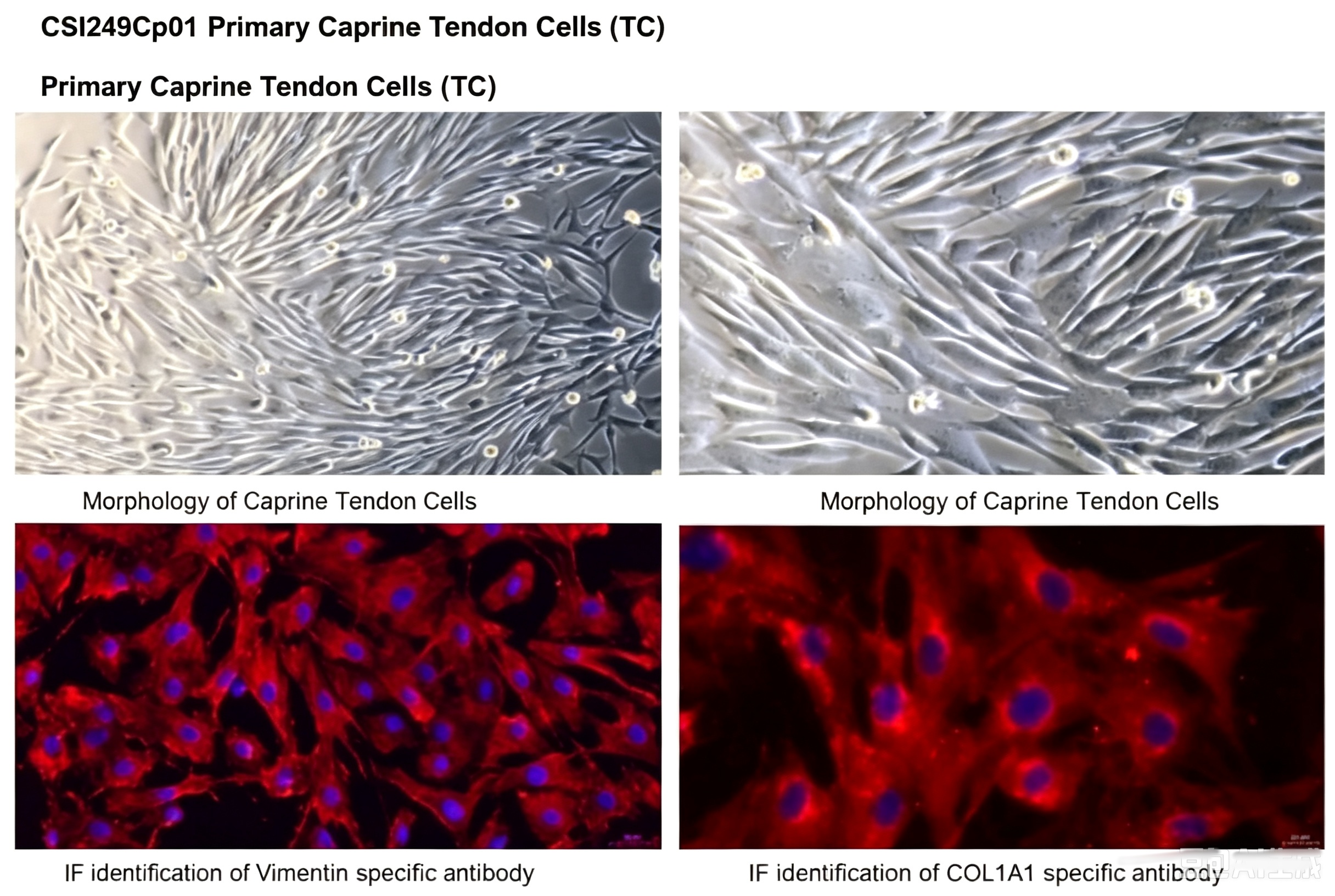
Peri-knee tendons including patellar and quadriceps tendons are essential structures sustaining joint stability and locomotor function. Tendon cells represent the major cellular population within tendon tissue, synthesising and maintaining highly ordered type-I-collagen-rich extracellular matrices. Tendon injuries such as patellar tendinopathy and tendon tears are highly prevalent in sports medicine, characterised by difficult healing and high recurrence rates. Primary tendon cells are core experimental tools for exploring tendon homeostasis, post-injury inflammatory responses, fibrotic mechanisms and mechanical-stimulation-dependent cellular functional changes, supporting tendinopathy and sports-medicine research.
Figure 5. Morphology and immunofluorescence identification of caprine primary tendon cells (Cat.No. CSI249Cp01)
Cloud-Clone: Source-Level Quality Control Establishes Reliable Starting-Points for Knee-Joint Cellular Research
Precision in knee-joint disease research builds upon the reliability of high-quality primary cells. Backed by self-operated SPF-grade animal facilities, ISO-certified quality-management systems and nearly twenty-years of cell-manufacturing experience, Cloud-Clone provides over 600 primary-cell products covering more than 10 species including human, mouse, rat, rabbit, dog, cat, sheep, guinea pig and chicken. For knee-joint-focused research, Cloud-Clone supplies core cell types including articular chondrocytes, synovial cells, synovial-derived mesenchymal stem cells, meniscus fibrochondrocytes and tendon cells. All cells are isolated from SPF-grade animal tissues and subjected to rigorous phenotypic characterisation and functional validation to guarantee high purity, high viability and batch-to-batch consistency. These cellular resources deliver solid support for mechanistic studies, drug screening and regenerative-medicine exploration targeting knee-joint diseases.
From cartilage to synovium, from meniscus to tendons: traceable, well-characterised primary joint cells lay solid foundations for every step of knee-joint-related research.
Unresolved translational gaps persist between laboratory cell assays and clinical outcomes for knee-joint-related disorders. Reliable primary-cell resources that faithfully mirror in-vivo physiology are indispensable to narrow such gaps. Cloud-Clone will keep expanding its knee-joint-relevant primary-cell portfolio, optimising source-level quality-control workflows, and empowering global researchers to unlock new insights for knee-joint disease mechanism research, therapeutic candidate screening and regenerative-medicine advancement.
About Cloud-Clone Corp.
Cloud-Clone Corp. is dedicated to the development and production of high-quality immunoassay reagents and detection solutions. With a focus on antibody engineering, multiplex assay development, and cross-platform compatibility, the company provides research tools designed to support precision medicine and advanced biomedical investigation globally. Our core products and services include the research and development of proteins, antibodies, ELISA kits, primary cells, and multiplex cytokine assay kits, as well as professional CRO services to fully meet the diverse needs of biomedical research and related fields.
For more information about Cloud-Clone Corp, visit www.cloud-clone.com.
CLOUD-CLONE CORP.(CCC)
Tel: 001-832-538-0970, 0086-27-8425-9552
Email: mail@cloud-clone.com, sales@cloud-clone.us
Visit us on social media:
LinkedIn: https://www.linkedin.com/company/cloud-clone-corp/
Instagram: https://www.instagram.com/cloudclonecorp2023/
TickTok: https://www.tiktok.com/@cloudclonecorp2026
SuKi Duan
CLOUD-CLONE CORP.WUHAN
email us here
Visit us on social media:
LinkedIn
Instagram
TikTok
X
Legal Disclaimer:
EIN Presswire provides this news content "as is" without warranty of any kind. We do not accept any responsibility or liability for the accuracy, content, images, videos, licenses, completeness, legality, or reliability of the information contained in this article. If you have any complaints or copyright issues related to this article, kindly contact the author above.
